In vitro means “in the glass”: The reactions take place outside the body. In vitro tests focus primarily on the reaction itself.
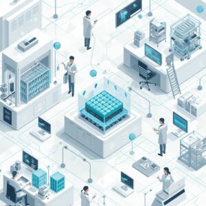
Advanced 3D in vitro methods are increasingly used throughout the drug development process (screening stages -techniques for identifying chemical compounds-, treatment optimisation).
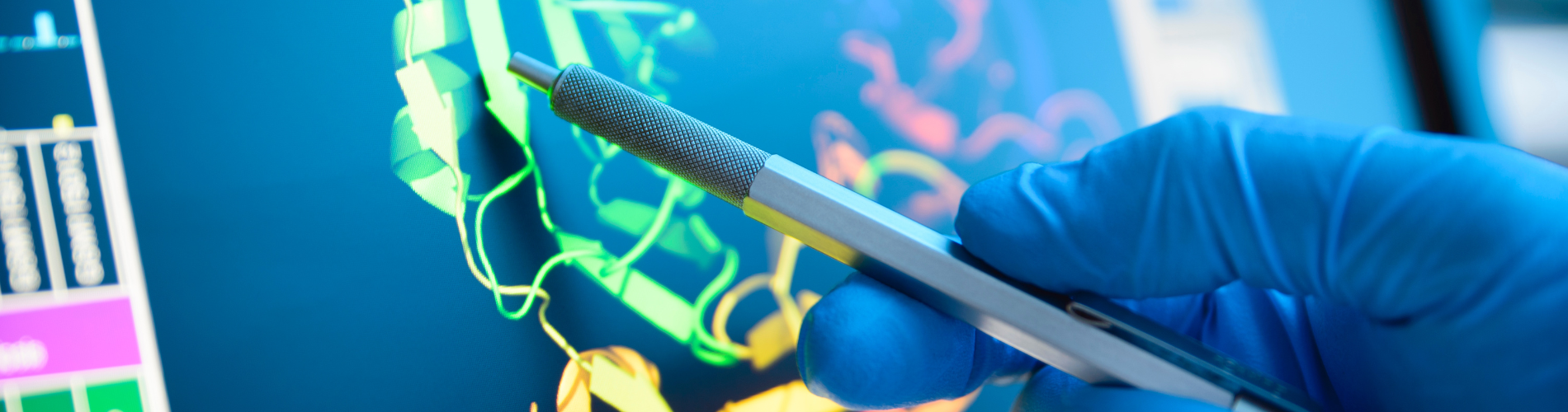
3D in vitro systems make possible to study a toxic mechanism of action and to use human cells, enabling the production of more human-relevant models.
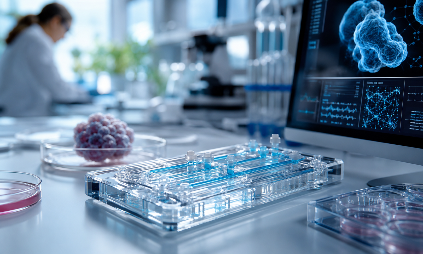
Microphysiological systems: Organs and organoids on-a-chip (O&OoC)
Microphysiological systems (MPS) are advanced 3D in vitro models that replicate the physiology of human organs and enable the culture of cells in a more natural, structured, and biologically relevant environment
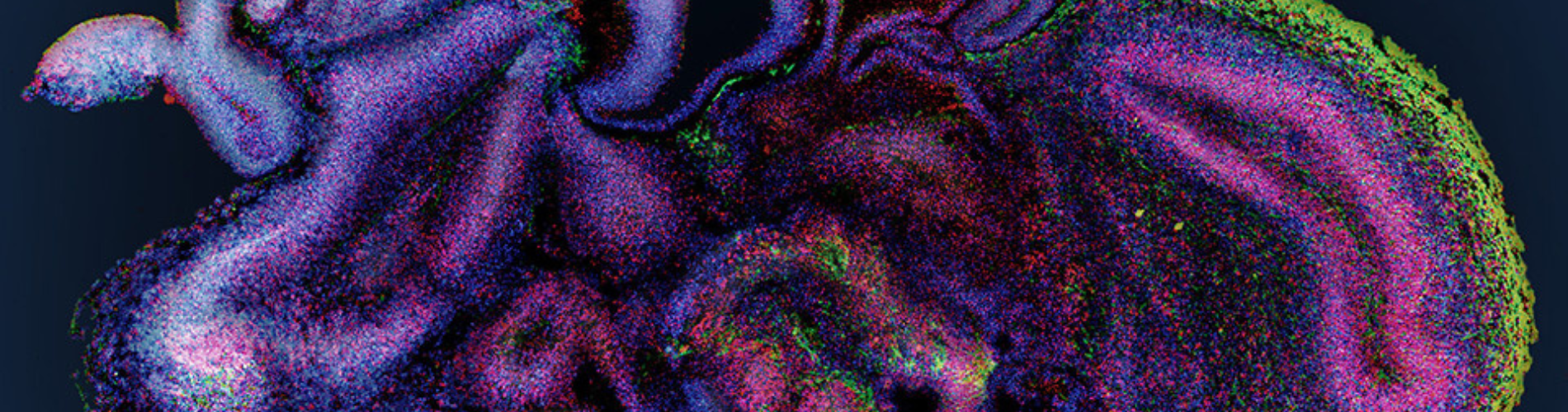
Organoids & Organs-on-Chip are Microphysiological Systems that make it possible to mimic the architecture and function of organs, and different functions of the human body.
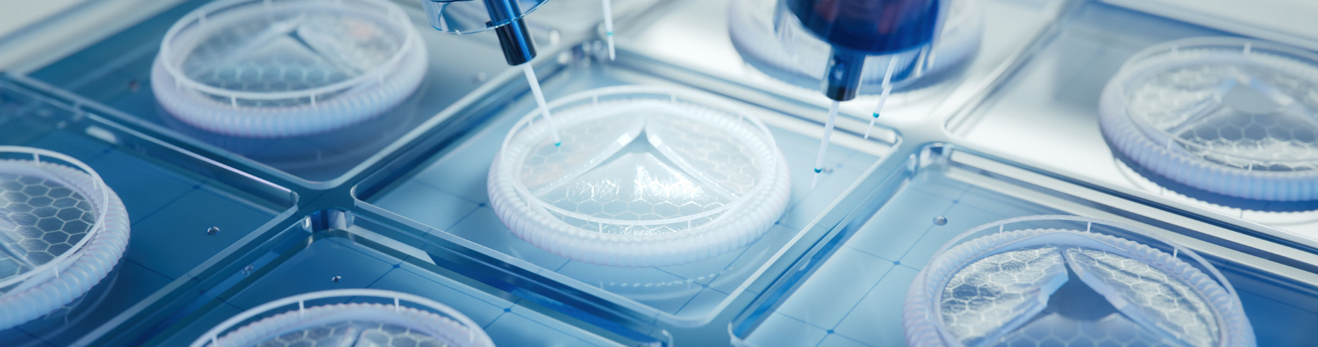
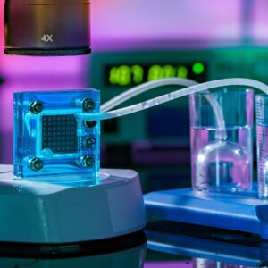
Organs-on-chip
OoC are small devices, basically the size of a computer memory stick, optically clear, usually made out of a flexible silicone rubber material. The device has tiny microchannels through which fluids flow, hence the name "microfluidics". From this, organ-level structures can be created by having two different tissue types interfaced across the membrane.
Learn more with Prof. Donald Ingber, pioneer of OoC technology, Wyss Institute, Harvard University

OoC technology aims to imitate the functions of an organ and the interactions between organs, in physiological or pathological conditions, through controlled biomechanical stimulations. With OoC, it is possible to study the propagation of a pathogen or test the effect of a drug in one or several lab-grown interconnected organs, using multi-organ chips.
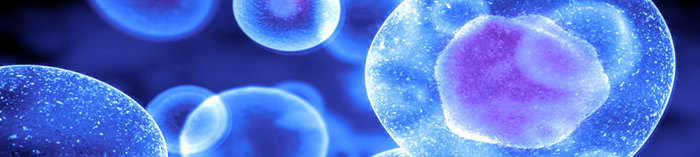
Organoids
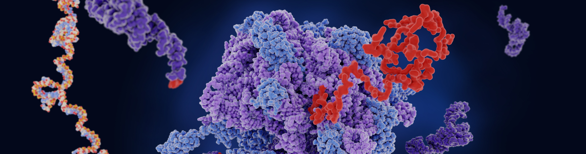
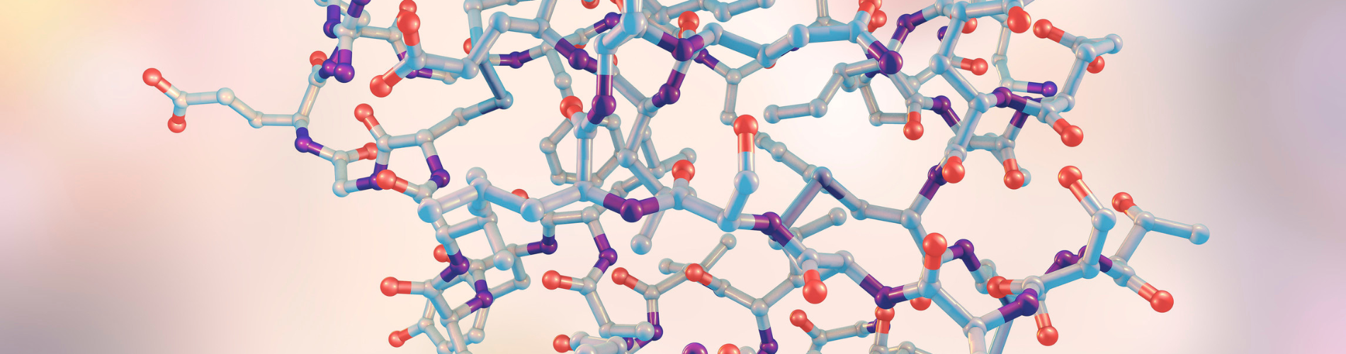
Organoids are 3D structure derived from pluripotent or multipotent stem cells1, progenitor cells that self-renew and self-organize through cell-cell and cell-matrix interactions reproducing, in vitro, certain architectural and functional aspects of native tissues.
Organoids mimic the architecture and function of the entire organ.
Examples of practical applications
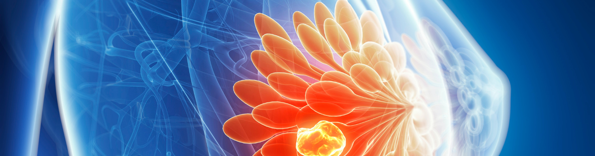
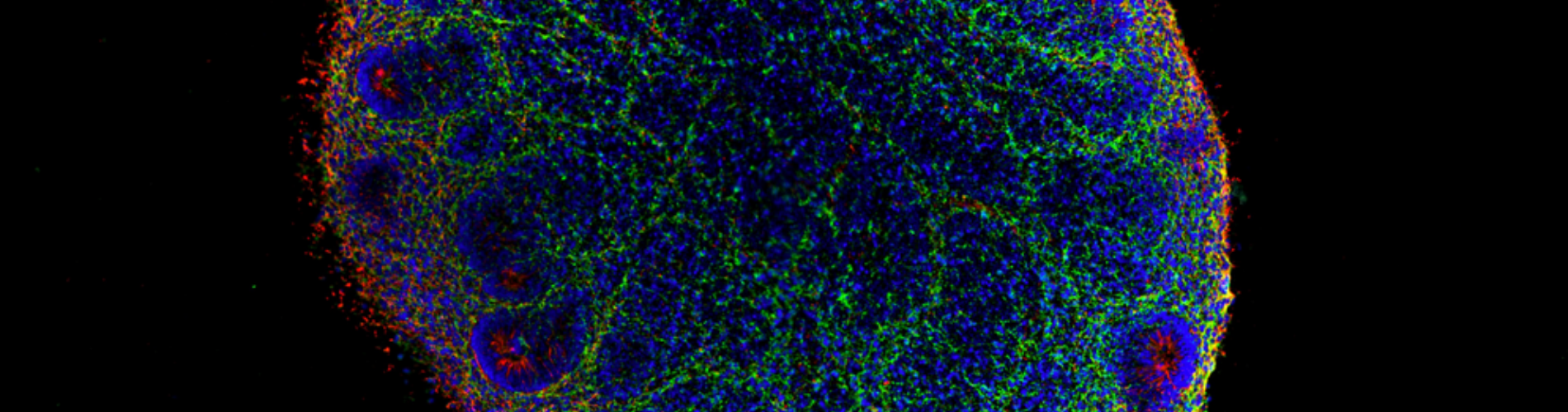
Tumor-on- chip to study cancerous tumors
The 3D glimpse project conducted by the FEMTO-ST Institute and Dr Agathe Figarol aims to create a tumor on-a-chip in order to better understand and treat glioblastoma, a very aggressive cancer with an average survival of one year after diagnosis. In this chip, the researchers seek to represent the tumor micro-environment via the use of different types of 3D-organised human cells in order to form micro-vessels. These will be infused to mimic blood flow in order to study the transport and effectiveness of new nano-drugs.
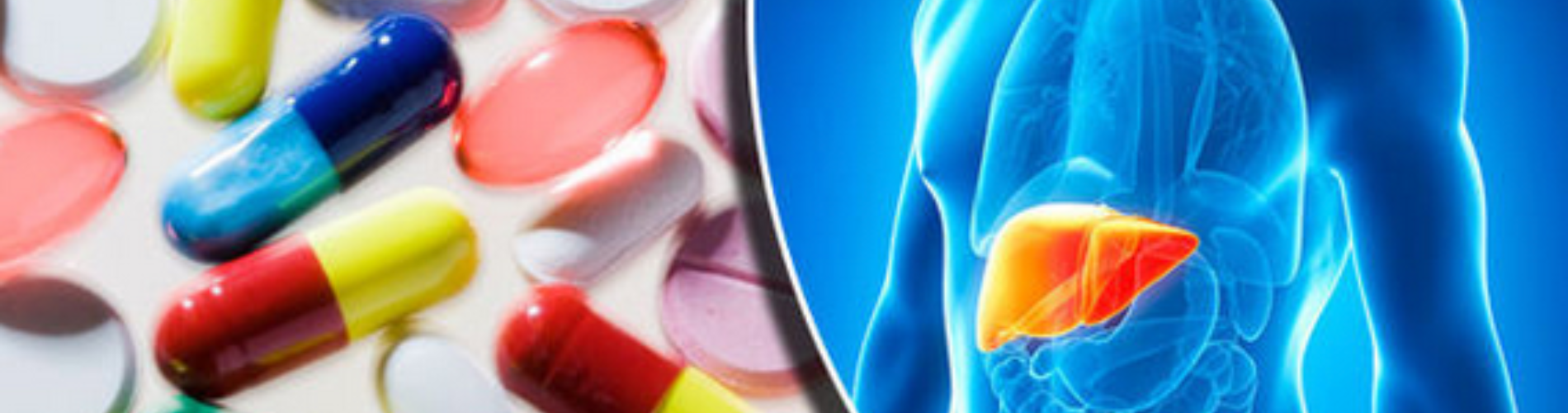
The liver-on-chip to analyze the toxicity of molecules for therapeutic purposes
The MimLiveronChip project is a biomimetic Liver-on-a-chip platform. Developed to recreate the analysis of liver metabolism and xenobiotic toxicity. MimLiveronChip seeks more particularly to explore the effects of the mechanical or biochemical microenvironment influencing the opening of the liver monolayer, in order to be able to generate it or, on the contrary, alter it.
Cell therapy
Cell therapy involves using iPSCs (induced pluripotent stem cells) or multipotent cells from the patient or a donor to graft cells to restore the function of a tissue or organ. The aim is to provide lasting treatment for the patient through a single injection of therapeutic cells.
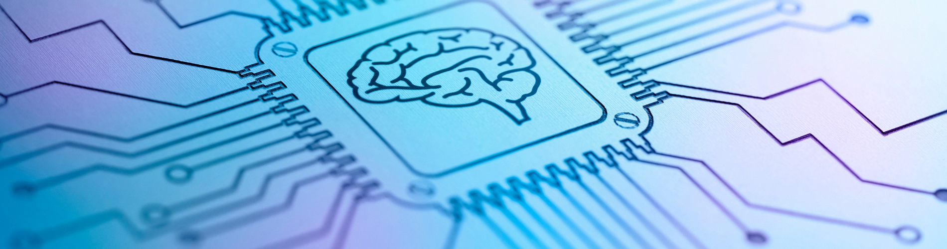
Organoid intelligence
Organoid intelligence (OI) is a new field of research in biological computing. Defined in 2023, it aims to develop a new form of computer, or "biocomputer", using 3D cultures of human brain cells (or brain organoids) and brain-machine interface technologies.
OI seeks to use lab-grown cerebral organoids to serve as "biological hardware." Scientists hope that such organoids can provide faster, more efficient, and more powerful computing power than regular silicon-based computing and AI while requiring only a fraction of the energy.
OI is also seen as a new frontier in biocomputing and biopharma drug discovery.