Alternative methods

Alternative methods


New Approach Methodologies and Non-Animal Technologies

Alternative methods are methods used in various scientific fields such as research and toxicity testing that do not rely on animal experiments.

Non-animal methods represent alternatives to the classic in vivo animal model.

The great originality of these methods is the development of knowledge specific to our species and/or to each individual by working on the different characteristics of human beings, which is impossible with the animal model.

The NAT (Non-Animal Technologies) database lists a wide variety of advances and studies related to non-animal methods.

In this article, learn more about the main areas of non-animal research, about the most advanced methods, and their applications.

Weekly news - France, Europe & World

Methods & non-animal technologies

In Vitro Methods

Clinical and applied research, toxicological tests.

Stem cells, Organs and Organoids on chip

Crédit: Laboratoire Poietis

Crédit: Laboratoire Poietis

In vitro means “in the glass”: The reactions take place outside the body. In vitro tests focus primarily on the reaction itself.

In vitro methods are increasingly used not only in the screening stages (techniques for identifying chemical compounds), but also during drug development. The ease with which they can be used, their isolation from any physiological context, which makes it possible to study a toxic mechanism of action, and above all the possibility of using human cells, which means that differences between species can be eliminated, make them an essential tool.

Stem cells

Crédit: Depositphotos

Crédit: Depositphotos

A stem cell is an undifferentiated cell capable of self-renewal, differentiation into other cell types and proliferation in culture.

Stem cells are most often derived from human surgical waste destined for incineration, such as cells found in skin and adipose tissue.

Technological advances have made it possible to programme these adult stem cells. They can give rise to virtually all the different types of cells in the body. They are pluripotent and known as iPSCs (induced pluripotent stem cells). Thanks to their properties, these cells can be used to regenerate or recreate destroyed tissues. Stem cells are used to design organs and organoids on a chip.


Organs and organoids on a chip
Crédit: iStock

Crédit: iStock

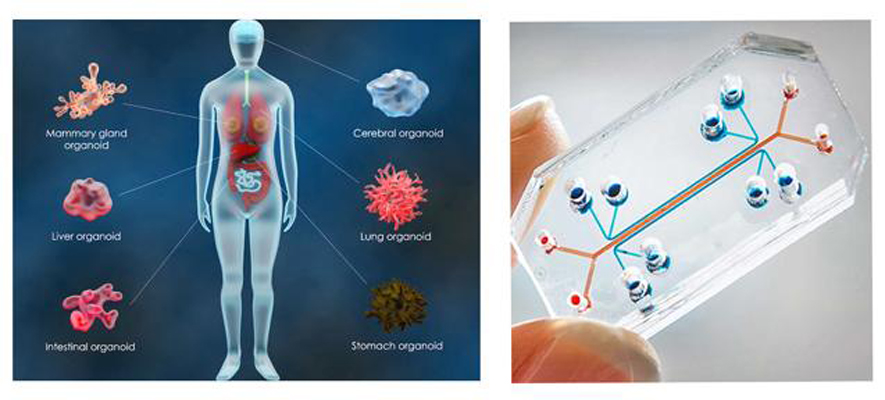
The aim of organoids or organs on a chip is to mimic the architecture and function of organs.

These devices are made up of human cells grown in a microenvironment, in valves punctuated by channels that can be connected using microfluidic technology.

It is designed so that the cells can simulate different functions of the human body.

With this technology, it is therefore possible to study the propagation of a pathogen or the diffusion of a drug in one or all of the organs cultured. It is also possible to simulate the interactions between organs using multi-organ chips.

Organoids are 3D cellular structures that mimic the architecture and function of the entire organ. They are obtained in particular from induced pluripotent stem cells (iPSC).

Clear advantages of in vitro methods over the use of animals

  • Accurately study the impact of the chemical reaction
  • Offer ease of cultivation and economic advantages for pharmaceutical industry production
  • Use human and not animal metabolism for toxicological studies

Some applications

Organoids on a chip to study cancerous tumors
Crédit: Depositphotos

Crédit: Depositphotos

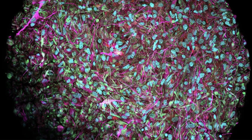
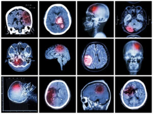
The 3D glimpse project aims to create a tumor on a chip in order to better understand and treat glioblastoma, a very aggressive cancer with an average survival of one year after diagnosis. In this chip, we will seek to represent the tumor micro-environment via the use of different types of human cells organized in 3D in order to form micro-vessels. These will be infused to mimic blood flow in order to study the transport and effectiveness of new nano-drugs.

The liver on a chip to analyze the toxicity of molecules for therapeutic purposes

The MimLiveronChip project is a biomimetic Liver-on-a-chip platform. Developed to recreate the analysis of liver metabolism and xenobiotic toxicity. MimLiveronChip seeks more particularly to explore the effects of the mechanical or biochemical microenvironment influencing the opening of the liver monolayer, in order to be able to generate it or, on the contrary, alter it.

Cell therapy
Crédit: Depositphotos

Crédit: Depositphotos

Cell therapy involves using iPSCs (induced pluripotent stem cells) or multipotent cells from the patient or a donor to graft cells to restore the function of a tissue or organ. The aim is to provide lasting treatment for the patient through a single injection of therapeutic cells. Cell therapy can also be used in the manufacture of organoids, for example.

In Omic Methods

Toxicological tests, clinical and fundamental research

Genomics, toxicogenomics, …

Crédit: Freepik

Crédit: Freepik

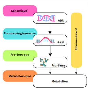
The terminology “in omic” or “omics” distinguishes more precisely the different cellular levels of analysis.

In omics methods are predictive tools that study the protein composition of cells and their activity via the occurrence of physiological, pharmacological or toxicological events.

Proteins have a wide range of functions within the cell and therefore contain a great deal of information about the state of the cell. They are essential components of the organism, determining how metabolism proceeds and which products are synthesised, broken down or degraded. The occurrence of a physiological, pharmacological or toxicological event will modify this protein composition and therefore the function of the cell.

Based on data from these in omic analyzes and comparison with control profiles or known characteristics, these methods help ensure patient safety during clinical studies or the marketing of drugs by providing better predictability and detection of adverse effects that may be observed in humans.


In omic methods represent four main levels of analysis:

Genomics

Genomics analyses the genome (all the genetic material of an individual or species), looking for altered genes or abnormal protein activities within a whole organism or organ.

Driven by advances in precision medicine, genomics is of particular interest in the study of cancers. It can provide invaluable information about the carcinogenesis of a healthy cell.

By understanding the genome of individuals, genomics is helping to develop a new form of personalized preventive medicine.

Transcriptogenomics or toxicogenomics

Transcriptogenomics (or toxicogenomics) studies changes in gene expression, from transcription to mRNA protein, in response to exposure to a chemical substance. As a reminder, a gene is a piece of DNA that stores all the information necessary for the proper functioning of organs and the body, and which is then transcribed into mRNA (messenger RNA).

Toxicogenomics is the result of the fusion of genomics and toxicology, and studies changes in gene expression in response to a chemical substance. Transcriptogenomics therefore aims to identify, classify and manage the latent and initial harmful and toxic effects of exposure to substances.

Proteomics

Proteomics involves the quantitative or qualitative analysis of all the proteins in an organism, biological fluid, tissue, cell or even cell compartment (also known as the proteome). Proteomics is often used as a complement to transcriptomics.

Metabolomics or Metabonomics

Metabolomics measures all the metabolites (small molecules) within an organ, tissue, cell or organelle, whether they originate from the body or the external environment.

Clear advantages of in omics over the use of animals

  • Early elimination of compounds with unacceptable toxicity through the identification of early toxicity biomarkers
  • Better prediction and detection of adverse drug reactions in humans
  • Prediction of the long-term effects of a chemical substance.
  • Contribution to the development of a better understanding of cellular mechanisms by enabling the analysis of several pathological pathways

Combining in omic methods with organoids to screen for the development of autism spectrum disorders

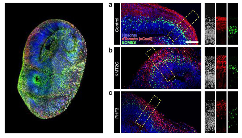
Researchers at the Institute of Molecular Biotechnology (IMBA) in Austria and the Swiss Federal Institute of Technology (ETH) in Zurich have come up with the idea of creating a brain organoid for early detection of the development of autistic spectrum disorders. The researchers are using genomics to analyse the genome (all the genetic material of an individual or species), looking for the 36 genes identified with autistic disorders and observing alterations and/or protein activity within the brain organoid as a function of the different genes expressed.

The system is called CRISPR-Human Organoids-Single-Cell RNA (CHOOSE) and makes it possible to visualise the genes involved in the autistic spectrum on the basis of 36 different genes identified.

Crédit: Knoblich Lab / IMBA-IMP Graphics / Li, C., Fleck, J.S., Martins-Costa, C. et al

Crédit: Knoblich Lab / IMBA-IMP Graphics / Li, C., Fleck, J.S., Martins-Costa, C. et al

In Silico Methods

Clinical, applied and fundamental research

3D and 4D bioprinting, organoid intelligence, etc.

The word silico is a derivative of the word silicon, a basic component of computers. In silico methods make it possible to predict the physicochemical and ecotoxicological properties of a substance from biomathematical models. These simulations provide an additional model to other methods and constitute a new type of scientific evidence.

The simulation model can be used during the initial phase of the drug development process, to test any hypotheses and modify the product to optimize its functioning or to estimate the probability that a given agent actually has the virtues of a drug, even before the first molecule is produced.

There are currently two categories of in silico technologies for conducting clinical trials: computational modeling and simulation, and artificial intelligence.

3D and 4D bioprinting

3D bioprinting uses a digital model to assemble and organize biological tissue components to artificially produce grafts or physiological models with the same properties as natural tissue. They are created layer by layer. In 4D, the aim is to develop the printed tissue over time.

Crédit: Laboratoire Poietis

Crédit: Laboratoire Poietis

Simulations of neurological mechanisms

Simulations of neurological mechanisms are computer models applied to the human brain. They have to deal with the very great complexity of neuronal interconnections. MRI scans of a patient's brain can now be used to develop neurological models. This technique opens up a wide range of possibilities for treating pathologies such as epilepsy.

Organoid intelligence

Organoid intelligence is a new field of research in biological computing. Defined in 2023, it aims to develop a new form of computer, or "biocomputer", based in particular on the performance of neurons. Researchers hope to make therapeutic advances through a better understanding of neurological functioning.

Crédit : Thomas Hartung, John Hopkins

Crédit : Thomas Hartung, John Hopkins

Definite advantages of in silico over using animals

  • Allow thousands of different simulation scenarios and virtual patients to be generated, enabling clinical studies on a much larger scale
  • Evaluate the specific performance of each patient, making it possible to identify the types of patient with the best response to treatment
  • Reduce the need for in vivo testing, enabling better planned clinical trials involving fewer patients

Some applications

Using 3D - 4D bioprinting to treat breast cancer

Crédit: Freepik

Crédit: Freepik

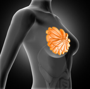
The Seno-Print project is seeking to improve breast reconstruction by developing 3D bioprinting of personalized biological prostheses. These bioprinted prostheses should adapt to the natural physiology of the breast, with the aim of limiting the number of additional interventions required to maintain and ensure the durability of the prosthesis.

Heart disease simulation

Crédit: Freepik

Crédit: Freepik

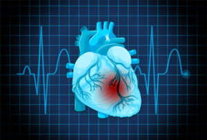
Computational analysis makes it possible to study the physical properties of blood circulation in the cardiovascular system via a fluid dynamics approach while adapting to many different cardiac pathologies. It is also possible to simulate the movement of the myocardial muscles. This approach makes it possible to predict clinical outcomes and assist in its design, support evidence of effectiveness, identify the most relevant patients to study and predict product safety.

Simulating neurological mechanisms to better guide epilepsy surgery

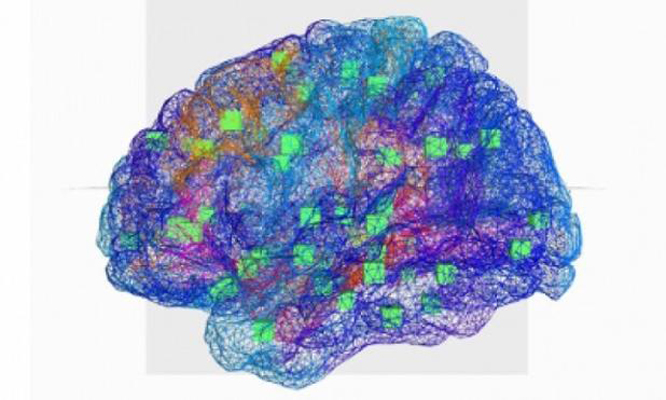
The aim of the Epinov project is both to improve analysis of the pre-surgical assessment of drug-resistant epilepsy and to better guide surgical strategies. To achieve this, Epinov is based on neuroinformatics brain simulation technology. The aim is to create a virtual brain in order to decipher seizures and improve epilepsy surgery by reproducing the abnormalities that cause epileptic seizures and providing carers with a model of the patient's epileptogenic zone.

Crédit: Epinov project

Crédit: Epinov project

Crédit: BodyInteract

Learning Methods

Clinical and surgical, human and veterinary training

Virtual simulation tables and interfaces, Synthetic human and animal models

Technological developments within education are gradually giving rise to new practices and training methods through simulation devices intended for both human and veterinary medicine students.

Indeed, medical training and surgical training in particular are based on learning theory and putting it into practice, in certain cases on animals, particularly pigs in surgery, before being followed by experience. clinical acquired through students' direct contact with patients.

In France in 2016, 34,000 animals were killed for educational purposes. In the same year, British universities and training centers used just 1,422 animals. In France, the number of animals used for teaching and training has increased by 31% since 2010.

Virtual simulation tables and interfaces

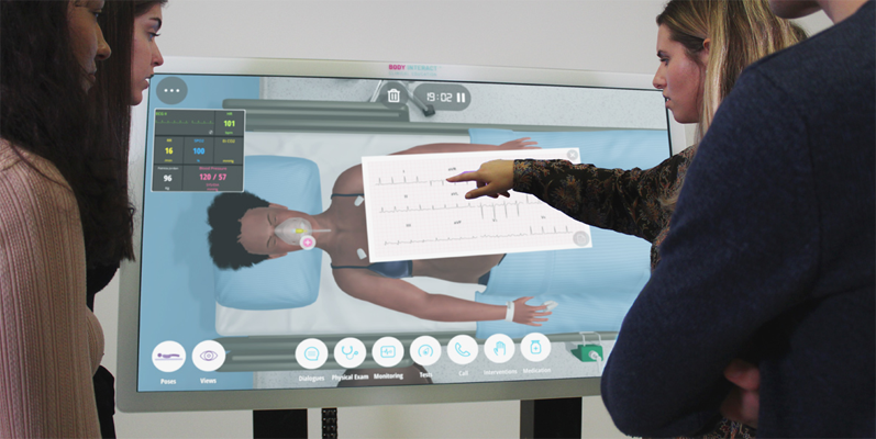
Virtual simulators and interfaces are used in the teaching of various clinical subjects such as neurology, cardiology, obstetrics, pediatrics, infectious diseases, etc.

Crédit: BodyInteract

Crédit: BodyInteract

The hyper-realistic virtual interfaces are obtained by fusing medical imaging data with anatomical data. They offer a veritable 3D digital anatomical library, enriched by collections of clinical scans and hundreds of scenarios to capture the complexity of real cases in a wide variety of environments.

These virtual patients with symptoms evolving over time based on physiological algorithms, provide relevant immersive simulation experiences representing a safe and interactive environment for the development of clinical reasoning and decision-making skills, as proposed by the BodyInteract device.

Synthetic human and animal models

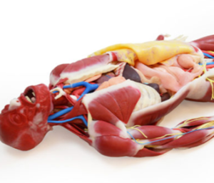
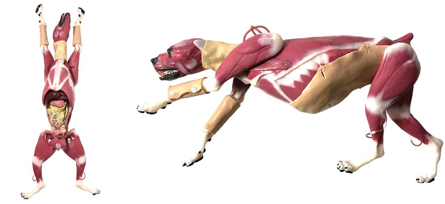
Synthetic models reproduce human and animal anatomy very accurately, including muscles, tendons, veins, arteries, nerves and individual organs. Manufactured from complex composites, the models produced reproduce the mechanical, physico-chemical and thermal properties of the living tissues concerned. The models can bleed and breathe, and use hundreds of replaceable muscles, bones, organs and vessels.

Crédit Syndaver

Crédit Syndaver

Work on this synthetic model technology was launched in 1993 at the University of Florida (USA). The materials and models developed since then can replace the use of animals in the study of medical devices, clinical training and surgical simulation, in both the human and veterinary fields, as well as in the evaluation of consumer products and ballistics testing, as proposed by Syndaver.

Crédit Syndaver

Crédit Syndaver

Clear advantages over using animals

  • Provide hands-on experience of dissections, manipulations and operations to understand the complexity of real cases, reducing the use of animals in learning
  • Provide a safe and interactive environment for the development of clinical reasoning and decision-making skills

In its resolution TA(2021)0387 adopted on 16 september 2021, the European Parliament considers that :

« the panoply of non-animal models of experimentation is growing and shows that it is possible to improve our understanding of diseases and speed up the discovery of effective treatments ».

The Parliament also points to bureaucratic obstacles that still stand in the way of acceptance of these methods, as well as problems with their use, which is not properly enforced, and insufficient funding for more effective development.

D’ding to the 2021 report (Dura, Gribaldo, Deceuninck), « Review of non-animal models in biomedical research — Neurodegenerative Diseases » of the Joint Research Center (JRC) of the European Commission :

“that heavy reliance on animal experimentation can hinder progress in certain areas of disease research”.